

When a short chain of amino acids hits a culture dish, the immediate question is rarely whether it looks promising on paper; the real question is whether the cells actually listen. In dermatological science, short-chain amino acid sequences engineered in the lab serve as precise molecular keys. Yet a massive gap exists between designing a sequence that mimics a natural collagen fragment and proving that it triggers measurable cellular behavior in a controlled laboratory setting.
That gap is where preclinical evaluation happens. Researchers rely on a tiered system of biological assays to trace how these molecules interact with cellular receptors, how deeply they penetrate tissue layers, and whether they genuinely alter gene expression or protein synthesis. Without this rigorous vetting, a peptide sequence remains an unverified hypothesis sitting in a lyophilized vial.
The Architecture of Modern Peptide Signaling
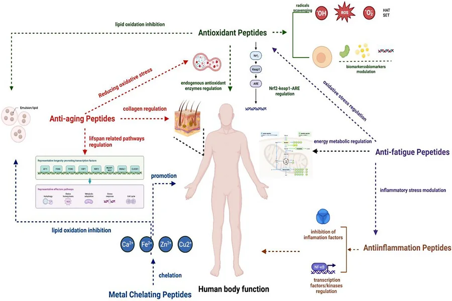
At the biochemical level, short amino acid sequences act as biological messengers. Rather than brute-forcing a structural change in tissue, they bind to specific cell-surface receptors or intracellular targets to initiate downstream signaling cascades. When scientists examine peptide signaling in the lab, they typically categorize these research compounds based on their primary mechanism of action.
Comparing signal peptides vs. carrier peptides highlights how differently these chains behave once introduced to a biological system. Signal peptides, such as palmitoyl pentapeptide-4, are designed to mimic fragmented extracellular matrix proteins; when fibroblasts detect these fragments, they interpret them as a sign of tissue breakdown and upregulate new collagen, elastin, and glycosaminoglycan synthesis. Carrier peptides, like copper tripeptide-1 (GHK-Cu), operate differently by stabilizing and transporting essential trace elements into the cellular environment, where those cofactors then activate enzymatic pathways involved in tissue remodeling and antioxidant defense.
Because sequence fidelity dictates receptor binding, sourcing and verifying raw materials is a foundational step before any biological assay begins. Cosmetic peptides are short, lab-engineered amino acid chains designed to mimic endogenous signaling fragments involved in structural protein synthesis and barrier maintenance. In laboratory settings, researchers investigate these compounds to map receptor affinity, cellular uptake, and downstream gene expression across controlled dermal models. Researchers looking to buy cosmetic peptides for laboratory assays typically verify batch-specific analytical data to ensure the sequences meet strict purity thresholds before introducing them to cell cultures. Securing well-characterized cosmetic peptides for research use prevents trace synthesis byproducts from skewing sensitive receptor-binding readouts.
Synthetic vs. Naturally Derived Peptides
Lab-engineered sequences offer a distinct experimental advantage over extracts harvested from botanical or animal sources. When evaluating synthetic vs. naturally derived peptides, reproducibility is the decisive factor. Naturally derived hydrolysates often contain a heterogeneous mix of chain lengths, residual salts, and undefined biological impurities that vary from harvest to harvest.
Solid-phase peptide synthesis eliminates that variability. Chemists assemble exact amino acid sequences one residue at a time, often adding N-terminal fatty acid chains like palmitoyl groups to improve lipophilicity and membrane permeability. High-performance liquid chromatography (HPLC) and mass spectrometry then confirm peptide purity and characterization, ensuring that any cellular response observed in a dish comes from the target sequence itself rather than an unknown contaminant.
How Are Synthetic Peptides Evaluated in Preclinical Skin Models?
Once purity is verified, the evaluation moves into living biological systems. To understand how are synthetic peptides evaluated in preclinical skin models, it helps to look at the progression from simple monolayer cultures to complex, multi-layered tissues. Each tier answers a different biological question, starting with basic cytotoxicity and moving toward functional barrier restoration.
2D Cell Culture vs. 3D Reconstructed Skin
The initial screening phase almost always begins with 2D cell culture vs. 3D reconstructed skin comparisons. In two-dimensional monolayers, researchers plate isolated human epidermal keratinocytes or dermal fibroblasts on flat plastic surfaces. These keratinocyte and fibroblast assays are fast, cost-effective, and ideal for high-throughput dose-response studies. Scientists can rapidly expose cells to logarithmic concentrations of a test peptide, measure cell viability via MTT or LDH assays, and quantify mRNA transcription changes using quantitative PCR.
Monolayers, however, lack spatial architecture. Cells grown flat on plastic do not form a stratum corneum, nor do they experience the paracrine crosstalk that naturally occurs between the epidermis and dermis. To bridge this gap, researchers transition promising candidates into 3D reconstructed human epidermis (RHE) or full-thickness skin equivalents. These 3D models feature stratified, differentiated keratinocyte layers exposed to an air-liquid interface, closely mimicking native tissue architecture and allowing scientists to test topical formulations rather than just aqueous solutions.
In Vitro vs. Ex Vivo Skin Models
Even the most advanced 3D reconstructed tissue lacks appendages like hair follicles, sweat glands, and a fully mature lipid matrix. That limitation brings researchers to the critical choice of in vitro vs. ex vivo skin models. While in vitro skin models rely on cultured cells or bioengineered tissues, ex vivo skin explants use viable human or porcine skin tissue obtained from surgical discards.
Ex vivo explants preserve the native extracellular matrix, intact resident immune cells, and complete stratum corneum lipid organization. Researchers mount these explants in Franz diffusion cells or culture them at an air-liquid interface to track both percutaneous absorption and biological activity simultaneously. A 2018 study published in the International Journal of Cosmetic Science demonstrated that certain lipopeptides exhibited markedly different penetration profiles and gene-upregulation kinetics in ex vivo human explants compared to monolayer fibroblast cultures, underscoring why intact tissue architecture alters experimental outcomes.
Measuring Repair: Assays and Functional Readouts
Demonstrating that a peptide enters a tissue layer is only half the work; researchers must also prove that it drives functional recovery. When designing experiments around tissue repair models and skin barrier models, scientists rely on a specific battery of physical and biochemical endpoints.
What assays measure skin barrier repair in the lab? Typically, investigators combine structural protein quantification with biophysical measurements of permeability.
- Scratch (wound closure) assay: Researchers create a standardized physical gap in a confluent monolayer of keratinocytes or fibroblasts, apply the test peptide, and use time-lapse microscopy to measure the rate of cellular migration and proliferation across the void over 24 to 72 hours.
- Transepidermal water loss (TEWL) in models: Using evaporimeters on 3D reconstructed skin or ex vivo explants after controlled barrier disruption (such as tape stripping or sodium dodecyl sulfate exposure), scientists track how quickly the tissue restores its water-holding capacity.
- Extracellular matrix markers: ELISA, Western blotting, and immunofluorescence staining quantify changes in pro-collagen type I, fibronectin, hyaluronic acid, and elastin production within the dermal compartment.
Alongside these primary endpoints, barrier function readouts often include transepithelial electrical resistance (TEER) to assess tight junction integrity, alongside histological staining for terminal differentiation markers like filaggrin, involucrin, and loricrin. When a synthetic sequence accelerates TEER recovery and upregulates filaggrin expression following chemical barrier disruption, researchers gain concrete evidence that the peptide modulates epidermal differentiation rather than merely hydrating the surface.
Interpreting Dose-Response and Stability Data
A common pitfall in preclinical peptide research is assuming that higher concentrations produce stronger biological effects. In reality, receptor-mediated peptide signaling frequently exhibits a bell-shaped or biphasic dose-response curve. At optimal nanomolar or low micromolar concentrations, synthetic peptides bind their target receptors and trigger robust transcription of repair proteins. Push the concentration too high, and receptors may undergo internalization and desensitization, causing the biological signal to plateau or drop off entirely.
Proteolytic stability further complicates data interpretation. Native skin tissue is rich in endogenous peptidases and proteases designed to degrade stray proteins. Unmodified linear peptides introduced into ex vivo explants or enzyme-rich culture media can be cleaved into inactive fragments within hours. Researchers routinely run parallel stability assays in skin homogenates to track the half-life of the intact sequence, checking whether chemical modifications like N-terminal acetylation, C-terminal amidation, or D-amino acid substitution successfully shield the peptide from enzymatic breakdown without compromising receptor affinity.
Translating Bench Data into Reliable Conclusions
Evaluating short-chain amino acids in the laboratory requires looking past single-assay spikes and examining the broader biological picture. A jump in collagen mRNA inside a 2D fibroblast monolayer means very little if the peptide cannot traverse the stratum corneum in an ex vivo explant or fails to improve barrier function readouts after physical disruption.
By pairing strict analytical verification with tiered biological testing, researchers separate genuinely bioactive sequences from inert ones. The most reliable preclinical datasets always connect the dots across scales: proving chemical identity first, mapping receptor-driven signaling in isolated cells, and finally confirming functional barrier and matrix restoration in intact, multi-layered tissue.